Body Thermography
Thermography or Digital Infrared Thermal Imaging, (DITI), is a totally non-invasive, painless procedure with no radiation and no contact with the body. DITI is a clinical imaging technique that records the thermal patterns of your body. Your thermal images are used by your healthcare practitioner to help diagnose and monitor pain or pathology in any part of your body.
THERMOGRAPHY CAN POTENTIALLY DETECT PROBLEMS IN THE BODY BEFORE THEY BECOME APPARENT CLINICALLY, OR SHOW SIGN AND SYMPTOMS.
Sheryl, owner of the Vista Natural Wellness Center and ACCT-Certified Clinical Thermographer, can discuss her own previous full-body thermographic results and how they indicted the presence of what was later determined to be a rare type of Non-Hodgkin’s Lymphoma.
Thermography’s major clinical value is in its high sensitivity to pathology in the vascular, muscular, neural and skeletal systems and as such can contribute to the pathogenesis, of this pathology and therefore, help the clinician make a diagnosis.
It is important to remember, that, “non-invasive” means NO radiation and NO contact with the body and is, therefore, totally safe, making it “the perfect procedure to be used as an annual screening test.”
Screening for illnesses utilizing Digital Infrared Thermal Imaging (DITI) or simply “Thermography” is available for both men and women. Due to its exquisite accuracy and sensitivity in detecting physiological pathology (not structural) in the human body, DITI has marked value in the early detection, diagnostic help, pathophysiology and the recovery process of disease.
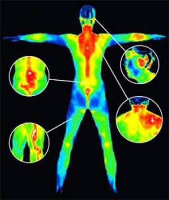
CLINICAL USES FOR DITI INCLUDE:
Inflammatory phenomena
- Infections
- Musculo-skeletal dysfunction
- Periosteal irritation
- Myofascial trigger points
- Active arthritis (joint disease)
- Sports injuries
- Monitoring the healing process
Vasomotor phenomena
- Radicular neuropathies
- Reflex Sympathetic Dystrophy (RDS)
- Neuropathological referrals
Vascular phenomena
- Angiogenesis (particularly perineoplastic growth)
- Varicies (thrombophlebitis and deep vein thrombosis (DVT)
- Ischemic phenomena
- Detecting sites of pathology BEFORE there is tumor formation
SOME OF THE MANY EXAMPLES FOR THE USE OF THERMOGRAPHY ARE:

BREAST PATHOLOGY
Thermography/DITI is an image of the physiology that is occurring in the body and not anatomical structure, DITI does not provide any of the same findings or information that mammogram or ultrasound provides, it is a different type of test. DITI shows information relating to vascular activity, inflammation, lymphatic activity, hormonal dysfunction and other ‘functional’ abnormalities. DITI is ideally placed as a screening tool to identify changes over time in the ‘early’ development stages, before there is more advanced pathology that can be detected with other tests. Mammogram and ultrasound shows ‘structure’, tissue densities can be evaluated, lumps can be measured, calcifications located and opinions given regarding pathology before biopsy ….. none of which DITI can provide.
Thermography/DITI was approved by the FDA in 1982 as an adjunctive screening test in addition to mammography for breast cancer.
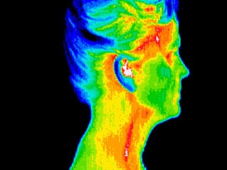
VASCULAR DISEASE PREVENTION
Thermography screenings can assess heart function and detect inflammation in the carotid arteries (which may be a precursor to stroke and/or blood clots). When inflammation and/or occlusion of the carotids are visible, your doctor may do additional testing. Earlier detection of a heart problem may save your life. Higher levels of heat in that region can also indicate immune dysfunction.
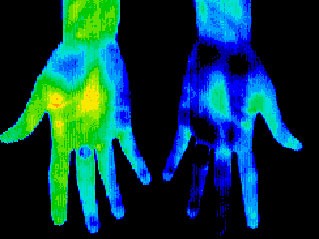
NERVE COMPRESSION / NEUROPATHY
Another aspect of thermal imaging which has gone largely unnoticed is in developing diabetic neuropathies of the feet, before the foot becomes insensate, by a 1-2 degrees centigrade drop in temperature as compared to the lower leg and usually the toes are not visible to the camera as they have become so hypothermic. This can occur several years before routine blood tests indicate diabetes, and as such, can give the patient time to treat the condition before permanent nerve damage occurs to the foot.
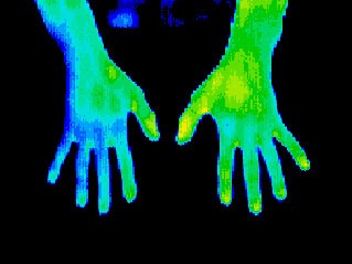
ARTHRITIS
Thermography can help you detect early signs of arthritis and differentiate between osteoarthritis and more severe forms like rheumatoid. Effective early treatment strategies can then be implemented, before you experience further degeneration.
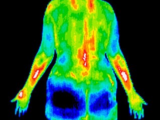
NECK AND BACK PAIN
Thermal pain patterns ‘light up’ white and red hot on a scan in the involved area. You can get relief faster and begin restorative care that more precisely targets the affected area.
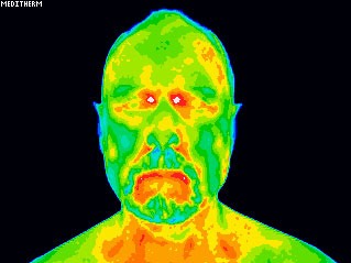
DENTAL ISSUES
If you have TMJ, gum disease or an infected tooth, this will show up on a thermal scan as white or red hot.
SINUS ISSUES & HEADACHES
Significant heat in your forehead or sinus region, revealed on a thermal scan, is an indicator that these areas in your body are inflamed and not functioning properly.

IMMUNE DYSFUNCTION
The immune system correlates to the T1 andT2 areas of your spine and high levels of heat in that region can indicate immune dysfunction. On the other hand, chronic fatigue, fibromyalgia and aching joints elsewhere in the body are just a few complaints that correlate to cool areas seen in this region.
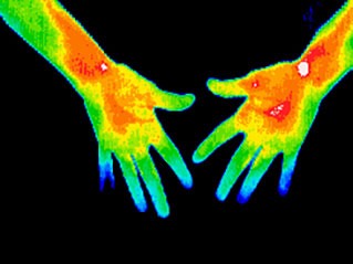
CARPAL TUNNEL SYNDROME (CTS)
This condition is often misdiagnosed. For instance, you may think you have CTS, yet the scan shows your neck is referring pain from a different affected area. This will help you receive the most appropriate treatment.
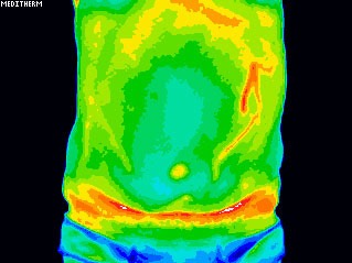
DIGESTIVE DISORDERS
Irritable bowel syndrome, diverticulitis and Crohn’s disease are often visible with thermography. If you’re able to address these conditions early on, you’ll find that restoring health is much more likely.
Other conditions that can be screened for with thermography include bursitis, herniated discs, ligament or muscle tear, lupus, nerve problems, whiplash, stroke screening, cancer, and many, many others.
- To define the extent of a lesion of which a diagnosis has previously been made;
- To localize an abnormal area not previously identified, so further diagnostic tests can be performed;
- To detect early lesions before they are clinically evident;
- To monitor the healing process before the patient is returned to work or training.
MEDICAL DITI IS ‘FILLING-THE-GAP’ IN CLINICAL DIAGNOSIS
- X rays, CAT Scans, Ultrasounds and MRIs etc., are tests of anatomy.
- EMG is a test of motor physiology.
- DITI is unique in its capability to show changes in physiology and metabolic processes.
- It has also proven to be a very useful complementary procedure to other diagnostic procedures.
We can image most of the entire body (brain is not able to be imaged due to the skull), the upper body from hips up, the lower body from hips down, or a specific region of interest such as the head, knees or breasts.
Medical uses of DITI have been used extensively in human medicine in the U.S.A., Europe and Asia for the past 30 years.

CONTACT US TO LEARN HOW THERMOGRAPHY (DITI) CAN BENEFIT YOU!
LEARN MORE ABOUT THERMOGRAPHY

JOIN THE VISTA COMMUNITY
Subscribe to our newsletter to learn more about our latest events, get healthy recipes and information to help you live a healthy lifestyle.
